In this new study, researchers compared three multiple imputation strategies for overcoming the missing discrete variable of gait speed in the Swedish National Study on Aging and Care (SNAC).
Aging Research
Read Crossref’s Top 10 Aging DOIs in 2023.

In a new study, researchers aimed to investigate the prognostic significance of senescence-related TME genes in head and neck squamous cell carcinoma (HNSCC) and their potential implications for immunotherapy response.

In a new study, researchers investigated the effectiveness and safety of EGFR-tyrosine kinase inhibitors in elderly patients with EGFR-mutated advanced non-small-cell lung cancer (NSCLC).

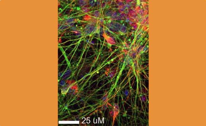
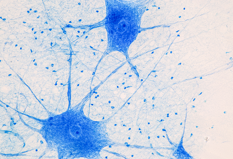
In a new editorial, researchers discuss interconnected mechanisms of neuronal functionality and available tools to investigate neuronal aging and longevity.

In this new study, researchers investigated the relationship between paternal age, the BEGAIN gene and autism.

In this new study, researchers revealed a novel role for LGR6 in enhancing WNT signals in pancreatic cancer.
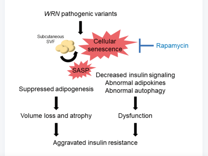
In this new study, researchers from Japan investigated the molecular mechanisms of subcutaneous fat dysfunction in Werner syndrome.